
| Optical imaging |
|
|
|
|
| RME Home |
| DCS Home |
| Research |
| Teaching |
| Publications |
| Contact |
|
|
| Email me |
|
|
|
|
|
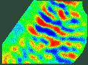
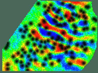
The reflectance of the mammalian visual cortex undergoes small changes (about one part in ten thousand) as its constituent neurons are activated. Monitoring the reflectance with a CCD camera allows detailed, simultaneous measurements of the active cortex to be made over tens of square mm.
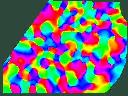
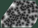
The picture shows an overlay of orientation pinwheels and ocular dominance columns in macaque V1 (right). The image is the superposition of ocular dominance columns (top left) and a distance map (bottom left) of each pixel from the nearest orientation pinwheel (middle left). There is a clear, but not perfect, correspondence between the location of pinwheels and the most monocular regions. (V2, where there are no ocular dominance columns, lies along the top left hand border of the images.)
The extremely small changes in cortical reflectance, elicited in response to a stimulus, sit in a background which fluctuates due to respiration heart beat and autonomous neural activity as well as instrument noise. We have developed sensitive methods for extracting the cortical signal are using them to investigate the structure of the visual cortex. The principal analytical tool is called the indicator function technique: these exploit the knowledge of when a stimulus was present and when it was absent to improve the signal to noise ratio over standard differential imaging methods. The method is explained in The separation, from background, of response evoked by stimulation (R.M. Everson, B.W. Knight & L. Sirovich, 1997. Biological Cybernetics. 77(6) 407-417). Current work with Alex Schmolck is focused on Bayesian extensions to this problem.
Spatial Frequency
We have used the indicator function technique to investigate the arrangement of neurons sensitive to spatial frequency. We find that the spatial frequency response is well approximated by admixtures of basis basis functions. Also, neurons sensitive to spatial frequency are, like orientation sensitive neurons, clustered in pinwheels . More details are given in Representation of Spatial Frequency and Orientation in the Cat Visual Cortex.
People
This work is a collaboration between many people at Mount Sinai Medical Center and the Rockefeller University in New York, particularly: Larry Sirovich, Udi Kaplan and Bruce Knight,
Papers
Here are some related papers.- R.M. Everson, A.K. Prashanth, B.W. Knight,
L. Sirovich & E. Kaplan, 1998.
Representation of Spatial Frequency and Orientation in the
Cat Visual Cortex. Proceedings of the National
Academy of Science USA. 95 8334-8338 Abstract.
- R.M. Everson, B.W. Knight & L. Sirovich, 1997. The separation, from background,
of response evoked by stimulation. Biological
Cybernetics. 77(6) 407-417. Abstract.
- R.M. Everson, E. Kaplan, B.W. Knight, E. O'Brien,
D. Orbach & L. Sirovich, 1997. Optical Imaging of the Mammalian
Cortex. In: Special issue of Biological
Bulletin, Advanced Computing for Biological Imaging. (To
appear.) Abstract.
- L. Sirovich, R.M. Everson, E. Kaplan, B.W. Knight, E. O'Brien & D. Orbach, 1996. Modeling the dynamics of the visual cortex. Physica D. Abstract.